The Neurobiology Of Impulsive Aggression
Aggression is a complex and heterogeneous construct. Mainly, we can distinguish between two types of aggression: premeditated (predatory, instrumental) and impulsive (affective, reactive) aggression. Here, we will focus on describing the neurobiology of impulsive aggression.
For the author Stahl, impulsive aggression can reflect “an emotional hypersensitivity and an exaggerated perception of threats. This may be linked to an imbalance between the top-down cortical inhibitory controls and the bottom-up limbic impulses ”.
Unrestricted impulsive aggression appears to be generally highly active in the amygdala area. In turn, it has little inhibitory activity in the orbitofrontal prefrontal cortex (COF) area. Conversely, when a person tries to control their impulsive aggression, the activity increases in COF. But the question is, where does aggressive behavior come from in the central nervous system?
Hypothalamus and periaqueductal gray matter
During the first half of the 20th century, studies were carried out in cats, specifically a region of the posterior hypothalamus was studied. Destroying this region was found to also “destroy” aggressive behavior of rage (false rage) that did not appear to be associated with actual anger. In addition, this behavior was not always directed towards the stimulus that had triggered it. When stimulated, this region provoked this angry behavior (2, 3).
Studies on the neurobiological bases of aggression in cats have led to the description of (4, 5):
- An affective attack, characterized by typical emotional responses of the sympathetic nervous system.
- A predatory attack, without these typical emotional responses.

The affective attack
This can be controlled from a large area of the medial hypothalamus. This extends to the brain stem where there are nerve centers that control the expression of the attack, such as hissing and grunting (6). In the affective attack, the following may also be involved:
- The medial tonsil. From it, the hypothalamus receives exciting information.
- The dorsal periaqueductal gray matter of the brainstem. The hypothalamus sends you exciting information. Furthermore, from this substance there are excitatory connections with the locus coeruleus and the solitary nucleus that mediate the autonomic responses during the affective attack (6).
The predatory attack
This type of attack is controlled by the brain from the lateral hypothalamus. Also from regions of the brain stem, such as the ventrolateral periaqueductal gray matter, among others. In addition, the lateral hypothalamus receives excitatory information from the central and lateral amygdala and inhibitory information from the medial amygdala. In this investigation, it is determined that both circuits inhibit each other. It happens that, while the cat is making a “predatory attack”, it cannot carry out an “affective attack” at the same time.
Haller (2014) states that the mechanisms described for the cat in the hypothalamus, periaqueductal gray matter and other centers such as the amygdala may also function similarly in humans. To this, the prefrontal cortex is added as a substrate for psychological factors.
Limbic structures (amygdala, hippocampal formation, septal area, prefrontal cortex, and cingulate gyrus) strongly modulate aggression through their connections with the medial and lateral hypothalamus (7).
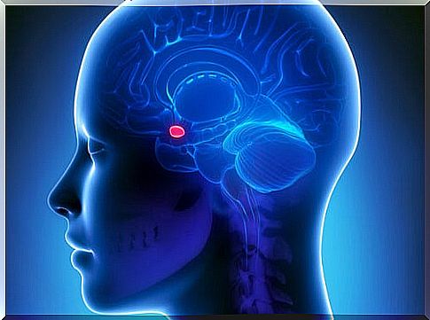
The amygdala
There appears to be a clear involvement of the amygdala in aggressive behavior. For example, in violent psychopathic subjects, a significant reduction in the volume of gray matter of the amygdala has been found in several studies (8, 9). However, other studies appear to show otherwise (1). What seems certain is that the amygdala plays a role in aggression. However, it is not very well known whether it increases or decreases in size when it occurs.
Regarding the activation of the amygdala, there have been studies in psychopaths that show lower levels of activity in the amygdala when they see violent images (1).
The prefrontal cortex in the neurobiology of aggression
What happens functionally in the PFC of violent subjects? A PET study carried out in predatory (psychopathic) and impulsive killers and in neurologically and behaviorally normal subjects (10) provided the following results in relation to the type of aggression and the activity of the prefrontal cortex:
- Impulsive killers had lower prefrontal activity and higher subcortical activity in the temporal lobe (which contains the amygdala) compared to control subjects.
- Predatory killers had prefrontal activity similar to that of control subjects but had excessive succortical activity.
It generally appears that violence causes at least one strange functional activity in the prefrontal cortex.
In general, studies on the neurobiology of aggression indicate subcortical structures such as the amygdala and other cortical structures as responsible for this behavior. It seems, therefore, that although the studies are not conclusive, they suggest that the violent behavior could be the result of a dysfunction in the cortical and subcortical activity.